Weber’s Syndrome: A Classic Midbrain Stroke Syndrome of Crossed Neurological Signs
Weber’s syndrome, also called Weber’s paralysis, is a rare but well-characterized midbrain stroke syndrome, typically caused by a lesion affecting the cerebral peduncle and the oculomotor nerve (cranial nerve III). It presents with a unique pattern of ipsilateral cranial nerve III palsy and contralateral hemiparesis, offering a textbook example of crossed (alternating) brainstem signs.
1. Historical Background and Eponym
Sir Hermann David Weber (1823–1918) was a German-born British physician. In 1863, he described a clinical syndrome combining third cranial nerve palsy on one side with motor weakness on the opposite side of the body. This led to the recognition of Weber’s syndrome as a distinct form of ventral midbrain lesion. Beyond neurology, Weber was a pioneer in tuberculosis treatment and a physician to several British prime ministers. He was knighted for his services to medicine in 1899.
2. Anatomy and Pathophysiology
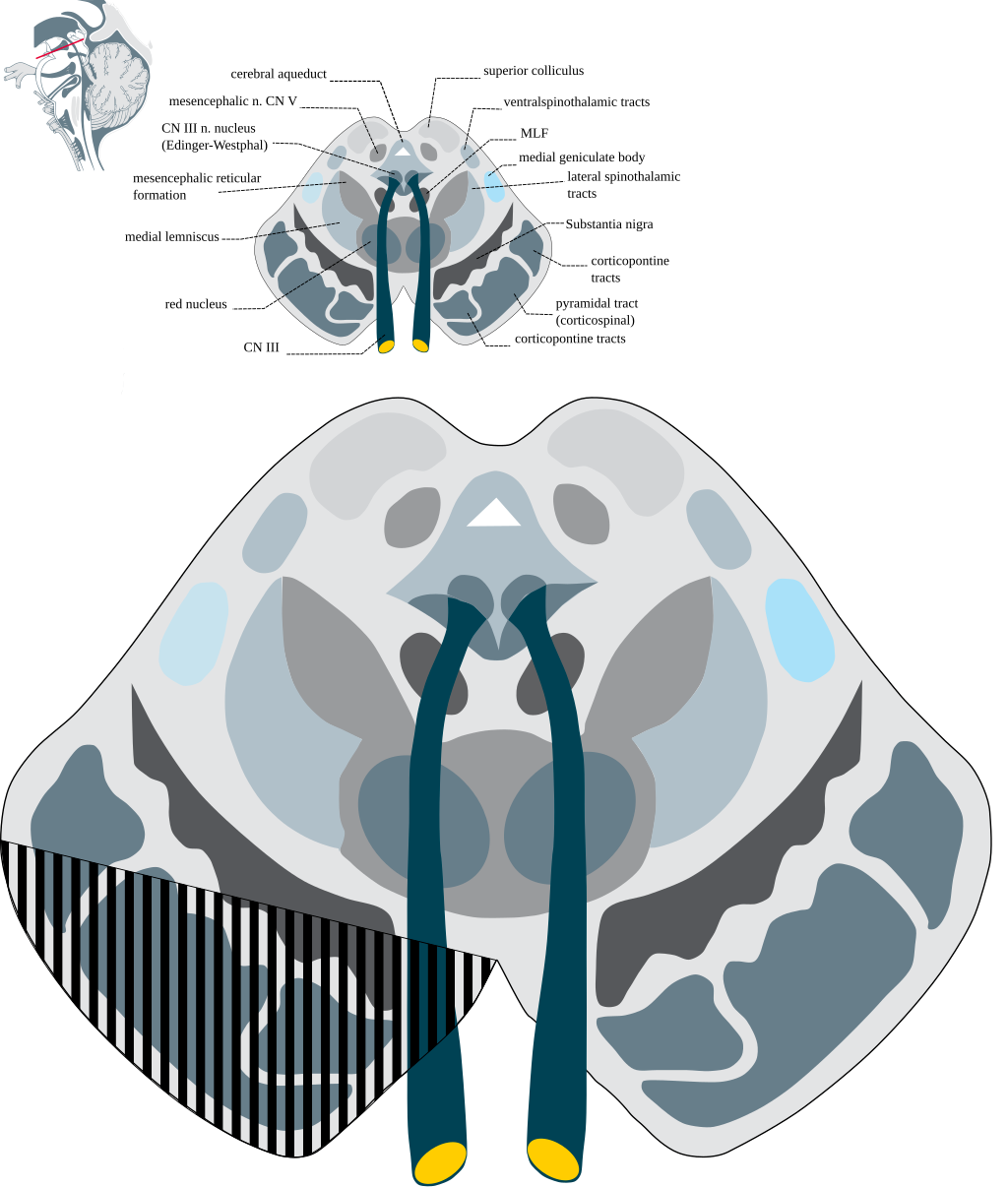
Weber’s syndrome results from a lesion in the ventromedial portion of the midbrain, particularly involving:
The oculomotor nerve (CN III) fascicles, which produce ipsilateral third nerve palsy
The corticospinal tract fibers in the cerebral peduncle, causing contralateral hemiparesis
Most common causes include:
Ischemic stroke, especially involving perforating branches of the posterior cerebral artery (PCA)
Hemorrhage, tumor, demyelinating disease, or trauma
3. Clinical Features
This crossed pattern—with cranial nerve deficit on one side and motor weakness on the other—is a hallmark of brainstem syndromes.
4. Differential Diagnosis
Weber’s syndrome must be differentiated from other midbrain syndromes with overlapping features:
5. Diagnosis
Diagnosis is based on:
Clinical neurological examination showing crossed cranial nerve and long tract findings
MRI of the brainstem to localize the lesion
Vascular imaging (MRA/CTA) to detect PCA branch occlusion or hemorrhage
6. Management and Prognosis
Acute Management
Ischemic causes: antiplatelet therapy, thrombolysis or thrombectomy (if indicated)
Hemorrhagic or structural causes: neurosurgical evaluation
Control of vascular risk factors (hypertension, diabetes, smoking)
Supportive Therapy
Physical and occupational therapy to restore strength and coordination
Ophthalmologic interventions for ptosis, diplopia, or persistent oculomotor palsy
Speech therapy if bulbar involvement occurs
Prognosis
Depends on the extent and cause of the lesion
Many patients experience partial recovery of cranial nerve III function
Motor deficits may improve significantly with early and sustained rehabilitation
7. Summary Table: Weber’s Syndrome Overview
8. Conclusion
Weber’s syndrome remains a classic example of brainstem neuroanatomy in action, where clinical signs precisely localize the lesion to the ventral midbrain. Thanks to the early insights of Sir Hermann Weber, this syndrome continues to serve as a valuable teaching tool and diagnostic clue in stroke neurology. Early recognition and intervention can lead to meaningful recovery in many patients.
Comment